Imaging and Treatment of Cancer through Combinations of Nanoparticles and Hormones
Carola Leuschner, Ph.D.
Page 3 of 4
In transmission electron microscopy images, you can see clusters of iron in lung cells, which are the metastatic cells in this case. Looking at and comparing the two images, i.e.
normal SPIONs, we hardly see any accumulation of iron in the tumor cells.
We can directly correspond the iron content in the lung tissue to the number of metastatic cells in these tissues. We receive a linear relationship proving that we can very specifically label the individual metastatic cells in the lung tissue. That was our first successful step in developing the contrast agent.
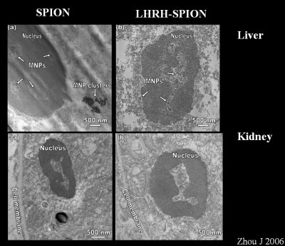
Image 7
We also needed to know if we had a large accumulation of nanoparticles in other organs
like the liver and kidney. Sure enough, we find that there's very little of the conjugated
nanoparticles in the liver and kidney, most of the free particles obviously accumulate in the liver.
The application of our study is in magnetic resonance imaging. In this case, we see magnetic resonance images from tumors of mice that have been injected with iron oxide nanoparticles from the experiment before. We can see already an increase in contrast, when we are looking at tumors from mice which have been injected with LHRH-SPIONs. In different MRI acquisitions and calculations, an increase in contrast compared to a normal contrast agent and a specifically targeted contrast agent.
Another important finding is, that these two images, which are Anisotropy Map images from mice which bear a tumor. The tumor is here from the same section.
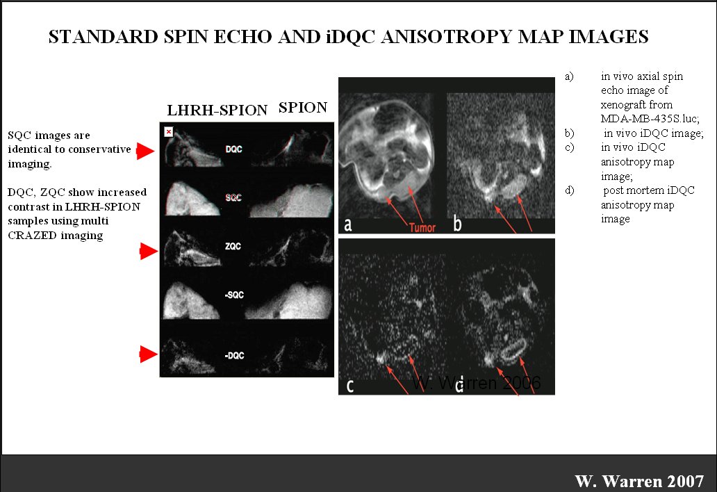
Image 8 [Click above for larger image]
If you look at the part marked by the red arrow, you see a lighting up of a small lump, and a lighting up of a circle, which is much sharper because this is a post mortem acquisition; we have a very sharp circle, and a very sharp lump to the right, both are areas of live tumor cells.
When we dissect the tumor, the center of the tumor always contains dead material, therefore unlikely to be labeled however; we can successfully label live tumor cells which are represented in the outer circle. This lump is very small, but it still contains exclusively live tumor cells. So the whole lump has accumulated the iron of the nanoparticles which can be imaged in magnetic resonance imaging.
This part of the article will demonstrate the preclinical development of any treatment, which is very similar to the previous approach, except that a drug is added to the iron oxide nanoparticles.
In order to cure cancers, we have to certainly reduce the 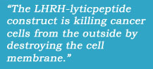 systemic toxicity. Also it's important to specifically target the diseased tissue, which has been shown in previous studies already that we have been able to target specifically the cancer cells by
putting LHRH on the surface of the nanoparticles.
systemic toxicity. Also it's important to specifically target the diseased tissue, which has been shown in previous studies already that we have been able to target specifically the cancer cells by
putting LHRH on the surface of the nanoparticles.
That also spares healthy tissue. Previously, we had shown a very small accumulation of free iron oxide nanoparticles in the peripheral healthy tissue. We have also shown that we can retain the targeted nanoparticles in the tumors and metastases.
And here we have conjugated the iron oxide nanoparticles with a drug, that is chemically a lytic peptide (Hecate), which is the same compound group as the LHRH. The LHRH-lyticpeptide construct is killing cancer cells from the outside by destroying the cell membrane. Here we have the same membrane and expose it to a lytic peptide. It's a peeling of the outer membrane of a cell, showing how a cell dies with that particular approach.
Obviously we had to ask ourselves the question if we construct a nanoparticle with a drug, is it still toxic or does it lose its toxicity by being processed and bound to the nanoparticle?
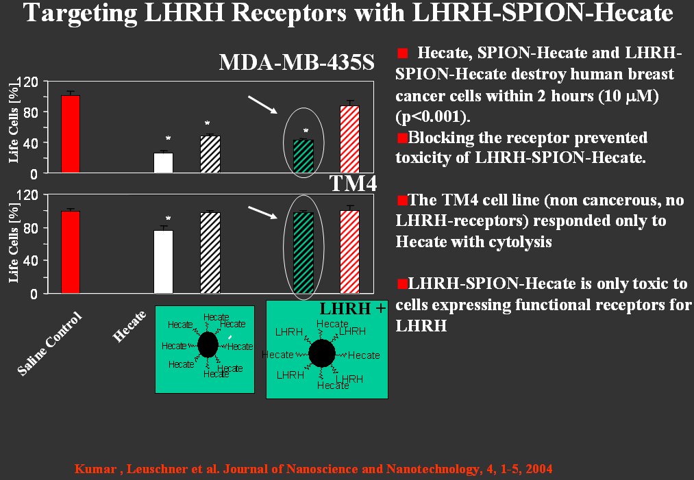
Image 9 [Click above for larger image]
In the in vitro experiment, we could show that we still have a very nice reduction of live cells; meaning this construct retains the toxicity of the cancer drug. It is very specific because the second cell line, the TM4 cell line, does not express the receptors. The cell line, therefore is not targeted, therefore it is not harmed.
It retains its viability, even under incubation of the cancer drug with the nanoparticle in comparison with the breast cancer cells, which has more than 60 percent cell reduction by incubation.
1 2 3 4 Next Page>